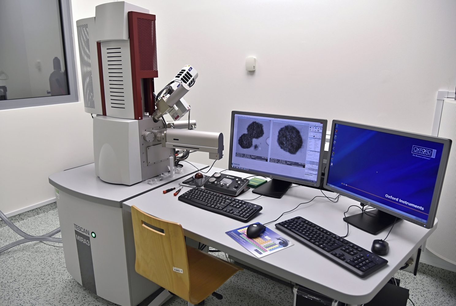
Scanning electron microscope (SEM) MIRA3 XMU
Scanning electron microscope (SEM) MIRA3 XMU Description • Scanning electron microscope (SEM) is used to study the morphology and topography of conductive and non-conductive materials in high resolution (micro to nano-scale). • Observation of surface samples with high depth of focus using multiple detection system (SE, BSE, STEM) including elemental analysis using energy dispersive spectrometer (EDS). Applications • Observation of both the surface and internal structure of micro and nano-objects (phase interface such as matrix-filler/reinforcement, particle distribution, aggregates and defects, fracture surfaces, porous 3D materials, units of supramolecular structure, etc.) • evaluation of the shape and dimensions (length, diameter, volume, roughness) of powders, tubes, short fibers • fast and highly accurate chemical microanalysis and elemental mapping of a sample surface • qualitative elemental analysis including determination of the distribution of each element • quantitative analysis of the individual elements in a sample • The structural analysis of polymeric materials, biopolymers and composites, biomaterials, ceramics, bones, teeth, substrates for tissue engineering, etc. Specification • High Brightness Schottky Emitter • Detectors: SE, BSE, In-beam SE, In-Beam BSE LVSTD STEM detector EDX analysis • High-vacuum (≤9x10⁻³Pa) or low-vacuum mode (7-500 Pa) • Magnification 25 to 1 000 000x • Acceleration voltage 200 V to 30 kV • X-Y-Z 130×130×100mm • Maximum specimen height: 106 mm
Specification
Scanning electron microscope (SEM) is used to study the morphology and topography of conductive and non-conductive materials in high resolution (micro to nano-scale). Observation of surface samples with high depth of focus using multiple detection systems (SE, BSE, STEM), including elemental analysis using an energy dispersive spectrometer (EDS).
Applications
• Observation of both the surface and internal structure of micro and nano-objects (phase interface such as matrix-filler/reinforcement, particle distribution, aggregates and defects, fracture surfaces, porous 3D materials, units of supramolecular structure, etc.)
• evaluation of the shape and dimensions (length, diameter, volume, roughness) of powders, tubes, or short fibers
• fast and highly accurate chemical microanalysis and elemental mapping of a sample surface
• qualitative elemental analysis, including determination of the distribution of each element
• quantitative analysis of the individual elements in a sample
• The structural analysis of polymeric materials, biopolymers and composites, biomaterials, ceramics, bones, teeth, substrates for tissue engineering, etc.
Technical parameters
| Cathode | High Brightness Schottky Emitter |
| Detectors | SE, BSE, In-beam SE, In-beam BSE, LVSTD, STEM, EDS |
| Vacuum modes |
High vacuum (≤9x10⁻³Pa) Low vacuum (7-500 Pa) |
| Magnification | 25-1 000 000x |
| Acceleration voltage | 200 V - 30 kV |
| X-Y-Z range | 130×130×100 mm |
| Maximum sample height | 106 mm |
Gallery
Publications
ZBONČÁK, M.; ONDREÁŠ, F.; UHLÍŘ, V.; LEPCIO, P.; MICHALIČKA, J.; JANČÁŘ, J., 2020: Translation of segment scale stiffening into macro scale reinforcement in polymer nanocomposites. POLYMER ENGINEERING AND SCIENCE 60 (3), p. 587 - 10, doi: 10.1002/pen.25317 (MIRA-STAN, CRYOGENIC)